Shared Equipment
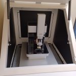
AFM Park NX10
Lab: OTC-305B
Park NX10 is a complete Atomic Force Microscope AFM system for general nanoscale imaging applications. It supports the following: Contact, non-contact and tapping mode. Electrostatic and magnetic force microscopy. The system is equipped with an acoustic enclosure with active vibration isolation. It uses the SmartScanTM operating software to produce high quality images. Automated or manual use, making initial training easier for multi-users.
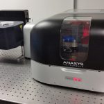
Atomic Force Microscope (AFM)/IR System – Anasys Instruments NanoIR2
Lab: R1-232
NanoIR2 system includes an Atomic Force Microscope (AFM) and a nanoIR2. It can be operated in contact and tapping mode for standard imaging. It can also be operated in advanced modes including force spectroscopy, conductive AFM, Kelvin Probe Force Microscopy, nanoinfrared spectroscopy, and nanothermal spectroscopy modes. The infrared laser covers the range 1500-1800 cm-1. Nanothermal mode covers the range 25 to 300C. Details on advanced mode are available during instrument training. The system is operated with Anasys software.
Atomic Layer Deposition (ALD) System – Ultratech Savannah S200
Lab: PVL-457
Savannah S200 ALD system supports thin film growth one layer at a time for 10s of nanometers at optimal scale. There are two modes of operation: Exposure Mode™, which allows conformal film growth at ultra‐high aspect ratio (i.e. > 2000:1) such as those found in porous foams, fibers, and nanogels; Continuous Mode™, which creates rapid growth of perfectly dense, uniform and conformal films. The unit is set up for aluminum oxide, titanium oxide, hafnium oxide, and zirconium oxide processes.
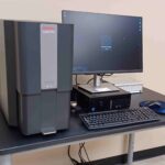
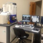
Benchtop SEM with EDS – ThermoFisher Scientific Phenom G6 ProX
Lab: OTC-305B
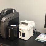
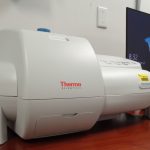
This benchtop SEM from ThermoFisher Scientific is optimized to be the ultimate all-in-one imaging system. It offers fast, high-resolution imaging featuring a fully integrated EDS detector. Light Optical Magnification ranges from 27x – 160 x and Electron Optical Magnification 160x – 350,000x. The resolution that can be obtained < 6 nm SED and < 8 nm BSD. The default acceleration voltages are 5 kV, 10 kV and 15 kV. The ability to run on high vacuum mode or low vacuum mode.
Confocal Microscope – Keyence BZ-X800
Lab: R1-216
The confocal microscope BZ-X800 captures images in the fluorescence, brightfield, and phase contrast fields for analysis (i.e. screening/quantification) and documentation. Its features also include XYZ stitching of large histology sections and time-lapse incubation for cell culture. The imaging system includes a built-in darkroom and allows sample analysis in slides, dishes, flasks, and microtiter plates. Some restrictions on lab use applies when using this microscope, contact the faculty in charge of this instrument (see "Instrument User Policy and Instrument List") for additional information.
Dynamic Light Scattering (DLS) & Laser Doppler Electrophoresis System- Malvern Zetasizer ZS90
Lab: PVL-466
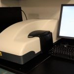
Zetasizer ZS90 measures particle size and zeta potential of particles or molecules in a liquid medium. Particle size measurement is in the range of 2nm to 3μm. The size range for zeta potential measurement is 3nm to 10μm. The unit is equipped with 633nm "red" laser for specific sample measurement.
Fluorescence Spectrometer – Horiba Nanolog FL3-11
Lab: OTC-305B
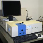
Nanolog FL3-11 has single-grating monochromator in the excitation and emission paths with PMT detector for use in routine fluorescence measurements. Right-angle or front-face emission collections are available for liquid and solid sample analysis.
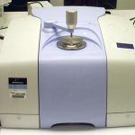
FT-IR Spectrometer – Shimadzu IRSpirit with QATR-S AT
Lab: PVL-466
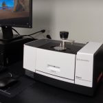
IRSpirit with QATR-S ATR is a compact FTIR spectrophotometer for qualitative and quantitative measurements in the 400-4000 cm-1 wavelength range. The single-reflection ATR measurement attachment (QATR-S) allows direct measurement of solid (i.e. powders, moldings, films, rubber) and liquid samples. LabSolutions IR software is used for instrument control/data processing and spectrum search is available through its database for material identification.
Integrated Confocal Raman and TERS Microscope – Horiba LabRAM HR Evolution Nano
Lab: OTC-305B
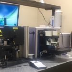
Horiba LabRAM HR Evolution Nano is an integrated confocal, tip-enhanced Raman system (TERS) and tip-enhanced photoluminescence (TEPL) microscope. It can be operated for (1) confocal Raman/PL spectroscopy and mapping (excitation wavelength available: 473nm, 532nm, 633nm, 785nm, 1064nm), and (2) Tip enhanced Raman/PL spectroscopy (TERS/TEPL) and mapping using integrated AFM (excitation wavelength available: 633nm, 785nm) for characterization of materials and defects with a resolution down to 10 nm. The integrated AFM options allow users to compare TERS and TEPL images with AFM topography, (photo)conductive, and Kelvin Probe Force Microscopy images for better understanding of the defects.
MicroPlate Reader – Tecan Infinite M200 Pro
Lab: PVL-447
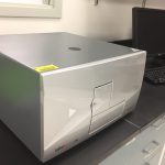
The Infinite M200 Pro can measure Fluorescence Intensity, Time-Resolved Fluorescence, Fluorescence Resonance Energy Transfer, Absorbance, Luminescence, and Bioluminescence Resonance Energy Transfer with wide range applications in analytical chemistry, cell biology, and protein science. Well plates and NanoQuant plate formats are available. Some restrictions on lab use applies, contact the faculty in charge of this instrument (see "Instrument User Policy and Instrument List") for additional information.
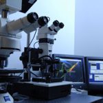
Optical Microscope – Olympus BX51M
Lab: PVL-466
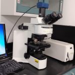
The BX51M Optical Microscope allows sample observation under 10X, 20X, 50X, 100X lens magnification on brightfield and darkfield modes. Eye and camera views are available; Jenoptik PROGRES GRYPHAX Camera provides live viewing and captures images for documentation/analysis.
Optical Microscope – Zeiss Axioscope 5
Lab: PVL-466
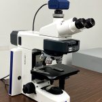
The Zeiss Axioscope 5 Optical Microscope is used for specimen imaging at objective lens magnifications of 5X, 10X, 20X, 50X and 100X. The LED illumination modes can be switched from reflected and transmitted lights. Imaging modes include bright-field and dark-field modes, and the user can observe the specimen with eyepiece or camera viewing. Axiocam 208 color camera provides live viewing and captures images for documentation/analysis.
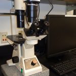
Phase Contrast Inverted Microscope – Olympus CK2
Lab: PVL-447
The CK2 Phase Contrast Inverted Microscope is equipped with Jenoptik GRYPHAX SUBRA 2.2MP Digital Scientific Camera to capture images for documentation and analysis. It uses ProgRes GRYPHAX Capture Software for live extended depth of focus, live tiling, basic measurements and annotations, multi-channel fluorescence overlay capabilities. Some restrictions on lab use applies, contact the faculty in charge of this instrument (see "Instrument User Policy and Instrument List") for additional information.
Scanning Electron Microscope (SEM)- Zeiss NVision 40
Lab: OTC-305B
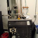
The NVision 40 can accommodate 2 specimens through the side air lock and 6 specimens through the main chamber for SEM magnification ranging from 30x - 900kx with resolutions of 1.1 nm @ 20kV and 2.5 nm @ 1kV. This is a used system and the actual resolution that can be obtained is around 10-20 nm. Detectors in place are EsB and BSE (in-column), SE (in-lens), and Everhart Thornley type SE detector (chamber). NVision 40 also has a NanoPattern Generating System (NPGS) which is capable of nanoscale E-beam Lithography. Applications include nanometer scale patterning and etching, as well as imaging.
Spinning Disk Confocal Microsope – Zeiss Axioskop 2 Mot Plus
Lab: PVL-448
Axioskop 2 Mot Plus Spinning Disk Confocal Microscope uses microscopy techniques ranging from transmitted light bright-field to epi-fluorescence together with image acquisition capability for image documentation and analysis (including 3D images) in the life science field. Some restrictions on lab use applies, contact the faculty in charge of this instrument (see "Instrument User Policy and Instrument List") for additional information.
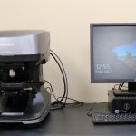
Surface Profiler (3D) – Keyence VK-X3000
Lab: OTC-305B
The 3D surface profiler VK-X3000 series offers both magnified observation and shape measurements with display resolution in height [z] (measurement range of 7 mm) and width [x/y] measurements up to 1 nm. It also possesses a measuring microscope that can analyze samples to quantify shape and roughness. The instrument employs two light sources: a short-wavelength laser source and a white light source. These two types of light sources enable acquisition of both laser intensity and color images, enabling the capture of fully focused and height/color map images. Film thickness measurements can be achieved from 80 μm (10x magnification) up to 3.5 μm (50x magnification) for transparent targets.
Thin Film Deposition System – AJA E-beam
Lab: PVL-442
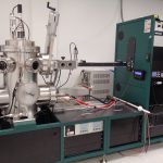
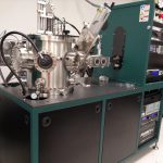
The AJA Electron Beam Evaporation System is an ATC-Orion-8E UHV system with 5 Pocket Linear E Beam Source on convenient slide mechanism for loading and service; it also features turbo-pumped vacuum load-lock. Contact the faculty in charge of this instrument (see "Instrument User Policy and Instrument List") for additional information.
Thin Film Deposition System – AJA Magnetron Sputterer
Lab: PVL-442
The AJA Magnetron Sputterer is an ATC-Orion 8 UHV with 5- 3" magnetron sputtering sources and turbo-pumped vacuum load lock with 6 position cassette. Contact the faculty in charge of this instrument (see "Instrument User Policy and Instrument List") for additional information.
Thin Film Deposition System – Thermionics E-beam & RDM Thermal Evaporator
Lab: PVL-438
The Thermionics e- beam evaporator applies intense electron beam to vaporize source material under high vacuum for deposition onto a desired substrate. It features 4 pockets for use with crucible liner size 7 cc. The RDM thermal evaporator applies low voltage (high amperage) for the vaporization process. There are 2 sources for use with boat size of 1 7/8 inch. The deposition system is a manual deposition up to 10-7 torr. Approved source materials for deposition are Au, Cr, Pd, Ni, Cu, Ti, Pt, Ag, In, Al, SiO2, Mo, and W – note that not all materials approved for the chamber will thermally evaporate due to high melting temperature.
UV-VIS Spectrophotometer – Evolution 220
Lab: PVL-466
Evolution 220 UV-Visible spectrometer is a state-of-the-art optical measurement device. It introduces the ISA-220 an Integrating Sphere Accessory (ISA). It operates in Reflectance mode and can easily be converted to operate in Transmittance mode. Both measurements are recorded using the INSIGHT software. The spectral measurement is in the wavelength range 200nm – 1100 nm.
Faculty Shared Equipment
FT-IR Spectrometer – Perkin Elmer Spectrum 100 Series
Lab: R1-232
The Spectrum 100 Spectrometer is a conventional FTIR spectrometer that can be operated in transmission or ATR configuration. The ATR accessory is recommended for analysis of solid and liquid samples. The range available for ATR is 650-4500 cm-1. The system is operated with Spectrum software.
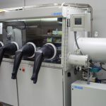
Glovebox/Thin Film Deposition System – MBraun MBEvap
Lab: PVL-457
The Glovebox/Thin Film Deposition System allows deposition of organic and metallic materials sensitive to oxygen and/or water by thermal evaporation or by solution-process using a spin-coater. Deposition procedure is carried out under inert atmosphere (i.e. N2). Some restrictions on equipment use applies, contact the faculty in charge of this instrument (see "Instrument User Policy and Instrument List") for additional information.
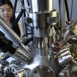
X-ray Photoelectron Spectrometer (XPS) – Fisons ESCALAB 2201-XL
Lab: PVL-438
The ESCALAB 2201-XL X-ray Photoelectron Spectrometer is a surface-specific instrument used to quantify the elemental composition and chemical states of samples. Its features include charge compensation for insulator samples, tilt control for angle-resolved XPS, and an argon ion etching gun for depth profiling. Thermo Avantage software is used for analysis on this instrument. Training is required instrument use - contact the faculty in charge of this instrument (see 'Instrument User Policy and Instrument List') for additional information.
